|
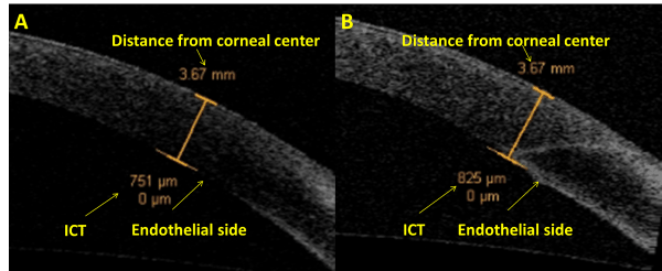
| Figure 2: Anterior segment optical coherence tomography images of incisional corneal thickness (ICT) at the endothelial side before the surgery (A) and at 3 months after the surgery (B) in the same eye. The two images also show the distance of the wound location from corneal center. |