|
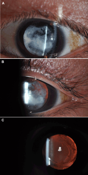
| Figure 4: Anterior segment photography. (A) A slit camera showed heterogeneous white cortical opacities of the left lens (mostly absorbed). (B) Partial fundus was visible through the dilated pupil. (C) Retro-illumination reveals the transparent intraocular lens after the phacoemulsification operation. |