|
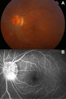
| Figure 6: Left fundus photography and fluorescein angiography after the phacoemulsification operation. (A) Fundus photography showing a leopard retina with a crescent surrounding the temporal part of the disc. (B) FFA (fundus fluorscein angiography) showing an unclear disc with a small amount of fluorescein leakage. |