|
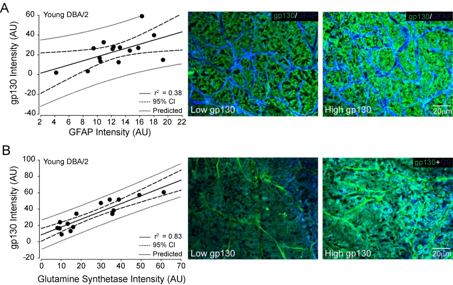
| Figure 10: GFAP and glutamine synthetase expression predict gp130 expression in the ganglion cell and nerve fiber layers of retina predisposed to glaucoma. Left panels: Regression graphs of fluorescent intensity for gp130 immunolabeling and intensities of GFAP (A) and glutamine synthetase immunolabeling (GS; B) in the ganglion cell and nerve fiber layers of midcentral to mid-peripheral regions of whole mounted retina from young DBA/2 mice. Data is plotted as intensity of gp130 (y-axis) versus intensity of GFAP (x-axis; A) and GS (x-axis; B) in arbitrary units. Solid line indicates regression line based on r2 value. Dashed line indicates the 95% confidence interval. Dotted line indicates predicted values based on r2 and 95% confidence. Right panels: Representative confocal micrographs depicting relationships between gp130 (green) and GFAP (blue; A) and GS immunolabeling (blue; B) labeling. For all, examples of low (left) and high (right) intensities are provided. Scaling is consistent for all images. |