|
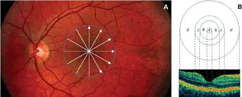
| Figure 2: Custom-built method showing macular sectors. A) Fundus image of a healthy eye showing the STRATUS OCT’s radial lines protocol. B) Regions shown are: foveola (a) with a diameter of 0.35 mm, foveal region (b) with a diameter of 1.85 mm, parafoveal region (c) with a diameter of 2.85 mm and perifoveal (d) region with a diameter of 5.85 mm. |