 |
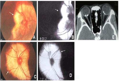
| Figure 1: Family 1, the first diagnosed patient was a 26-year-old female. A. Color image of the right fundus revealed the osteoma lesion at the posterior area (arrow). B. Right eye FFA revealed the osteoma lesion, which exhibited a strong fluorescence intensity (arrow). C. Color image of the left fundus revealed the osteoma lesion at the posterior area (arrow). D. Left eye FFA revealed the osteoma lesion, which exhibited a strong fluorescence intensity (arrow). E. Bilateral orbital CT scanning revealed high density areas of osteoma lesion in the posteriors of both eye rings. |