 |
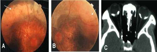
| Figure 2: Family 1, the older sister of the patient was 30 years old. A. Color image of the right fundus revealed rear atrophy and the osteoma lesions (arrow). B. Color image of the left fundus revealed rear atrophy and the osteoma lesions (arrow). C. Bilateral orbital CT scanning revealed high density areas of osteoma lesion in the posteriors of both eye rings. |