|
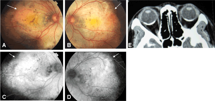
| Figure 3: Family 2, the first diagnosed patient was a 22-year-old female. A. Color image of the right fundus revealed the osteoma lesion at the posterior area (arrow). B. Color image of the left fundus revealed the osteoma lesion at the posterior area (arrow). C. Right eye FFA revealed the osteoma lesion, which exhibited a strong fluorescence intensity (arrow). D. Left eye FFA revealed the osteoma lesion, which exhibited a strong fluorescence intensity (arrow). E. Bilateral orbital CT scanning revealed high density areas of osteoma lesion in the posteriors of both eye rings. |