|
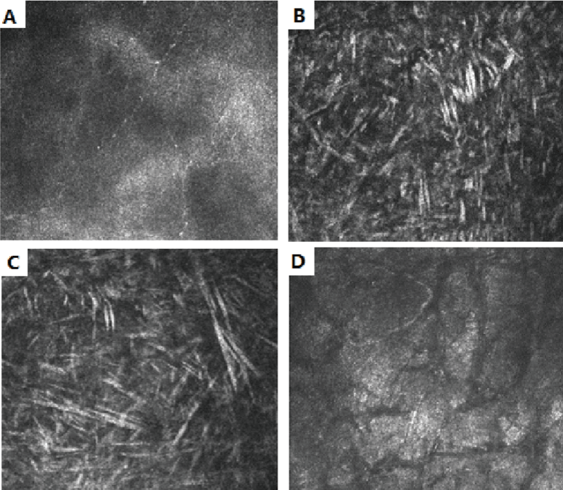
| Figure 3: (A) In vivo laser confocal microscopic images of the translucent basal/subepithelial nerves. (B and C) Brightly reflective crystalline materials were found in the anterior and mid stroma. (D) Crack-like striaes were observed in the posterior stroma. |