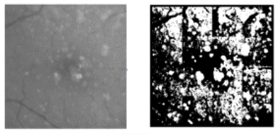
Figure 4:
Retinal cropped image showing drusen, vessel and background (left) and mask showing region of interest with removing vessel and partial background (right).