|
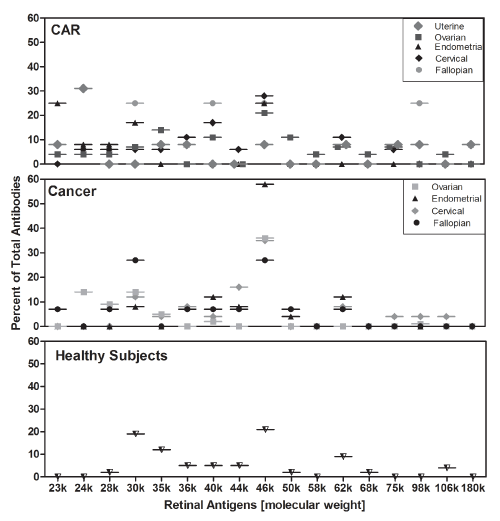
| Figure 2: Distribution of specific retinal autoantibodies according to the retinal proteins targeted by autoantibodies within each group of patients. X-axis shows retinal proteins marked by their molecular weight (k=1000). Graph represents the percent of antibodies reacting with each protein within a group. |