|
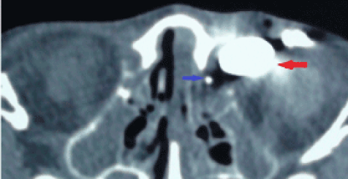
| Figure 1D: Axial computed tomography: Injection of contrast material from orificium fistula and normal lacrimal puncta (first patient). Red arrow: lacrimal diverticulum filled with contrast material; Blue arrow: normal lacrimal sac filled with contrast material. |