|
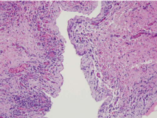
| Figure 2D: Haematoxylin-eosin stains revealed a cystic cavity lined by pseudostratified columnar and cuboidal epithelium with interspersed lymphocytes, plasmacytes and granular leukocytes. The wall of the mass also consisted of fibrous connective tissue (×200) (second patient). |