|
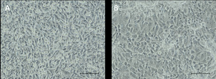
| Figure 1: Phase-contrast photomicrograph of OCM1 (A) and C918 (B) UM cell lines, growing as a monolayer in 25 cm2 flasks (see text). Magnification: 200×. Details of cell morphology showing a larger mean size with prevalence of spherical elements in C918 and spindle-like elements in OCM1. |