 |
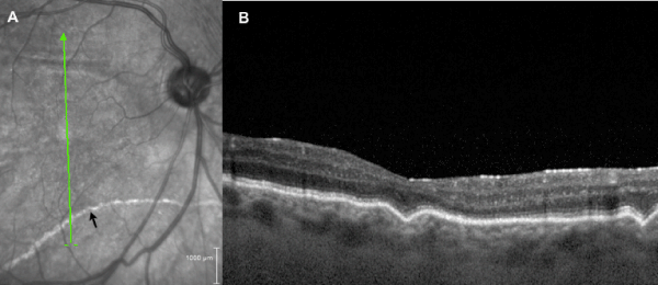
| Figure 3: Chorioretinal folds following scleral buckle. (A) Scanning laser ophthalmoscopic image demonstrating subretinal proliferative vitreoretinopathy (arrow) and scan position for SD-OCT image in (B) demonstrating chorioretinal folds involving the choriocapillaris, RPE, and outer retina; this 43 year-old patient with a spherical equivalent of -1.25 diopters has a normal subfoveal choroidal thickness of 223 μm. |