|
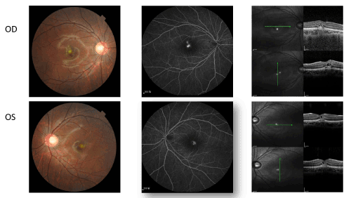
| Figure 1: Fundus photographs (left) , fluorescein angiogram (middle) and OCT-images (right). 2 days after the laser pointer beam exposure, fundus photographs showed yellowish-grey spot without hemorrhage in the foveal region, the OCT showed intraneurosenory edema and disruption of the RPE layer at foveal central region, fluorescein angiogram showed ill-definded leakage in the foveal region. |