 |
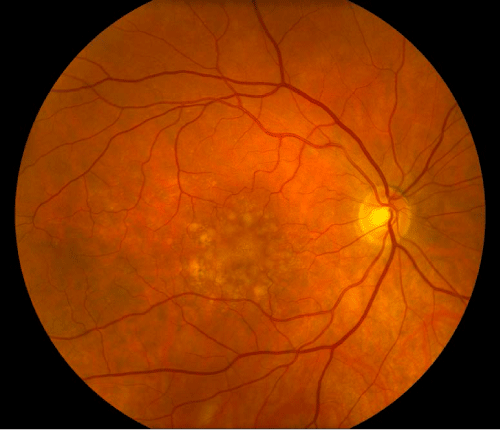
| Figure 1: Color fundus photo of high-risk non-exudative agerelated macular degeneration. There are numerous large (greater than 125 micrometer) soft drusen with central confluence in the fovea. Additionally, areas of RPE pigmentation abnormalities can be noted in the areas outside the drusen confluence. |