|
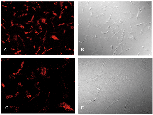
| Figure 2: Dil-labeled LDL staining of cultured porcine (A, B) and human (C, D) TM cells. A, C: Cells were stained with Dil-labeled LDL and visualized with a confocal fluorescent microscope after 2-3 passages. B, D: Normarski view of the same cells. Original magnification × 400. |