|
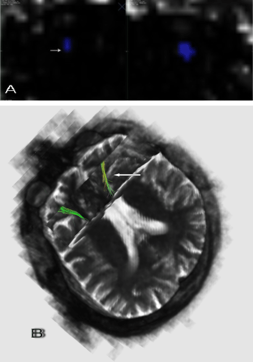
| Figure 3: Imaging of glaucomatous neurodegeneration in optic nerves. (A) Temporal nerve fiber loss can be seen in the severely affected eye (arrows) in b0 coronal plane images of an asymmetrical patient (B) Isotrophy and irregularity (arrows) in the optic nerve tractographies in the severely affected eye. |