|
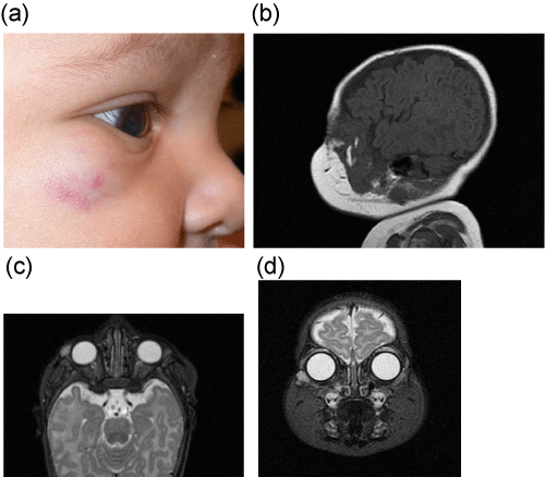
| Figure 4: A) Subcutaneous capillary hemangioma with deep bluish hue and soft borders. B) Well-circumscribed, lobulated mass with serpiginous flow voids at the infero-lateral orbital rim on T1- weighted MRI, sagittal view. C,D) The lobulated lesion has thin septae and slight hyperintensity on T2-weighted sequences (axial, coronal views). |