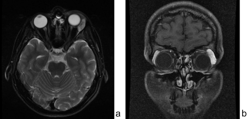
Figure 2:
(a) Axial T
2
weighted MR image with fat suppression and (b) coronal T
1
weighted post contrast MR image with fat suppression showing tumor mass in the left lacrimal gland area.