 |
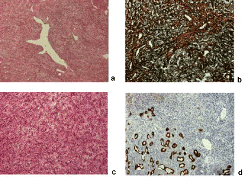
| Figure 5: (a) Hemangiopericytoma - "staghorn" configuration of vessels [HE, 60x], (b) typical arrangement in reticulum stain [silver impregnation, 100x] (c) section of the tumor with a predominance of small blood vessels [HE, 100x] (d) residual structures of the lacrimal gland inside the tumor [immunohistochemistry Cytokeratin AE1/3, 100x]. |