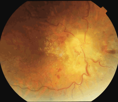
Figure 1b:
Fundus Picture showing established papilledema in the form of disc edema, peripapillary haemorrhages, venous dilatation & tortuosity with few peripapillary exudates.