 |
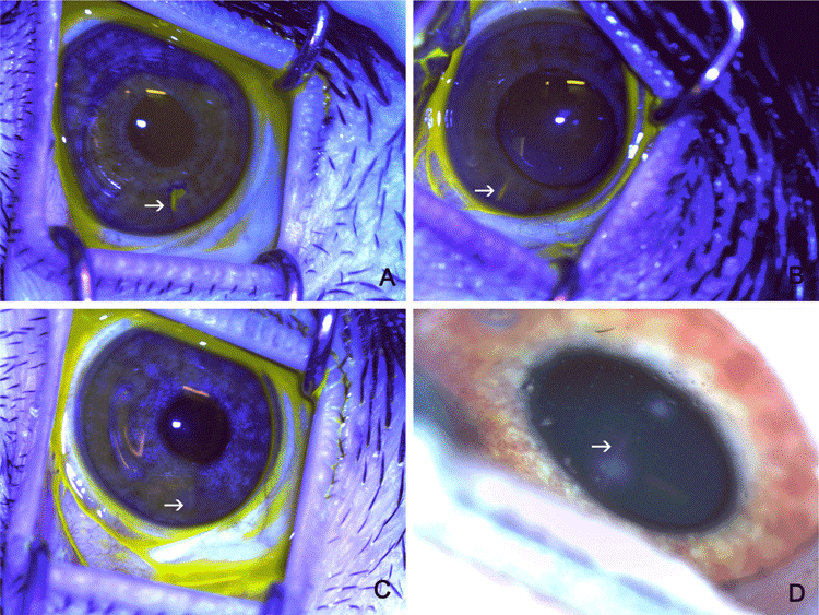
| Figure 3: Time-dependent changes in the morphology of the incision (arrow: incision location). (A) Staining by fluorescein at 24 hours showed a diffuse incision site. (B) At 1-2 days, the incision site was shaped like a fish mouth. (C) On day 3, the incision site was linear. (D) At 3 months after insertion, the wound site appeared as a linear haze. Panels A-C, fluorescein staining. Panel D, clinical photograph. |