|
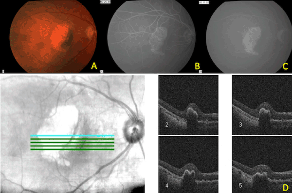
| Figure 2: Patient 1: Post-Treatment OD. A: Colour photo showing the scrolled RPE and the choroid visible in the void of RPE, B: Early FA showing the choroidal circulation visible through the RPE defect, C: Late FA showing the congruous RPE defect with no staining, D: OCT images through the RPE tear: The abrupt edge of the RPE is clearly visible in the cuts labeled 2 and 3. |