 |
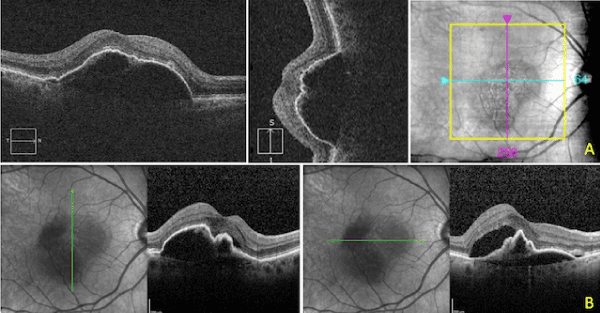
| Figure 3: Patient 2: Before RPE tear. A: A large PED OD is visible two months prior to the tear. The patient was treated at this visit, B: There is increased subretinal and intraretinal fluid, and the PED takes on an irregular shape. RPE strain is visible in the horizontal cut as irregular peaking of the RPE. The patient was treated again at this visit. |