|
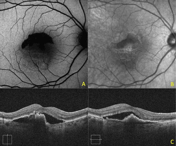
| Figure 4: Patient 2: After treatment. A: Autofluorescence photo OD demonstrating the striking absence of RPE, B: Red-Free photo of the same area, the corrugated RPE is visible inferior to the torn area, C: OCT through the RPE tear showing the retracted and corrugated RPE. |