 |
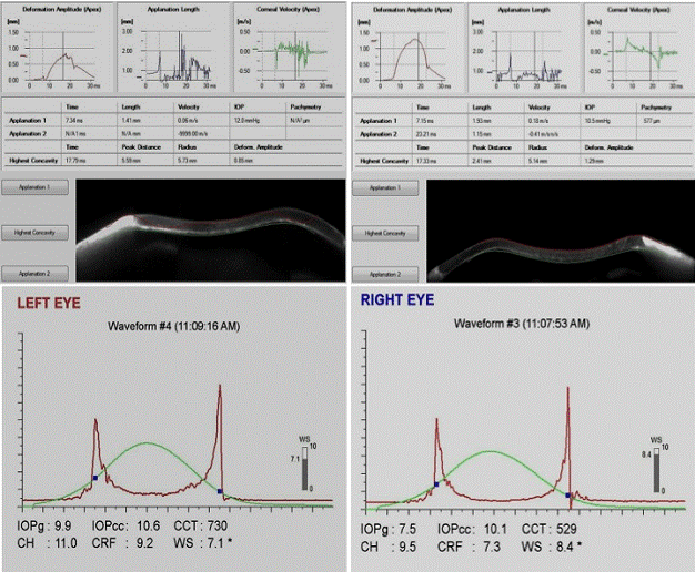
| Figure 2: Images showing the corneal biomechanical profiles using the Corvisc ST (upper left (for left eye) and upper right (for right eye) respectively) and the waveforms picked up using the ORA (bottom left (for left eye) and bottom right (for right eye) respectively) for sibling 1. |