|
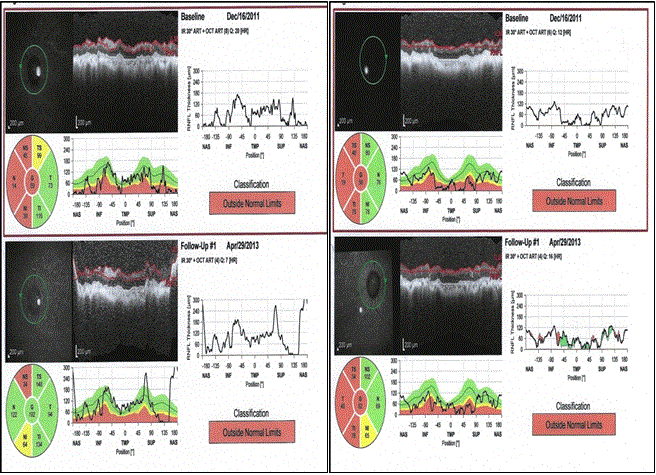
| Figure 7: Retinal nerve fibre layer (RNFL) scans (left – left eye and right –right eye) done for sibling 2 using the spectral domain- optical coherence tomography. It shows thinning of RNFL secondary to glaucomatous optic neuropathy and optic atrophy. |