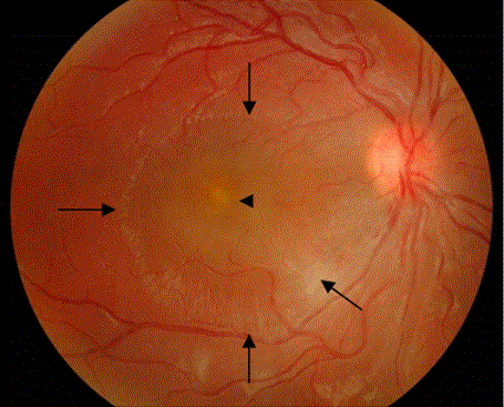
Figure 3A:
Fundus photo of patient 11 showing small foveal infiltrate (arrow head) and significant macular edema (long arrows) (before treatment).