 |
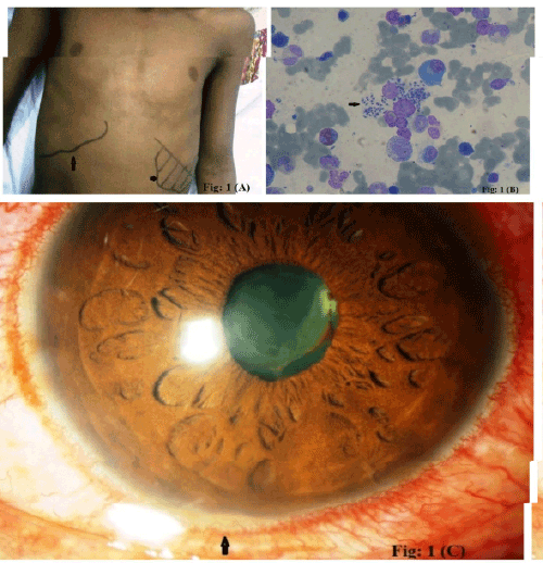
| Figure 1: Showing clinical as well as microscopic evidence of Kala azar first relapse along with post Kala azar Uveitis. Figure 1a showing hepatomegaly and splenomegaly (arrow marks)], Figure 1b showing Giemsa stained LD bodies (arrow mark) in bone marrow smear], Figure 1c showing post Kala azar anterior uveitis with fibrinous exudates (arrow mark) after treatment with liposomal Amphotericin B. |