 |
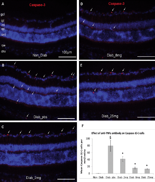
| Figure 3: Prevention of caspase-3 (+) cells by anti-TNFα treatment. The STZ-induced diabetic mice were IVT injected antibody biweekly for 3 months. (A-E) The sample images of activated caspase-3 staining for non-diabetic (A), diabetic with the treatments of PBS (B), 1 μg (C), 5 μg (D) and 10 μg (E). (F) Quantitative results were expressed as mean ± SD (n=10). $p<0.05 compared to non-diabetes. *p <0.05 compared to diabetes with PBS treatment. Scale bar: 100 μm. |