 |
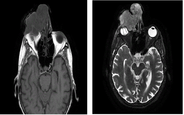
| Figure 3:MRI shows a left orbital mass (left) transaxial T1 weighted MRI image demonstrates an 8 cm mass that is hypointense relative to cerebral cortex with peripheral contrast enhancement. (Right) transaxial T2-weighted MRI the mass is isointense relative to cerebral cortex. |