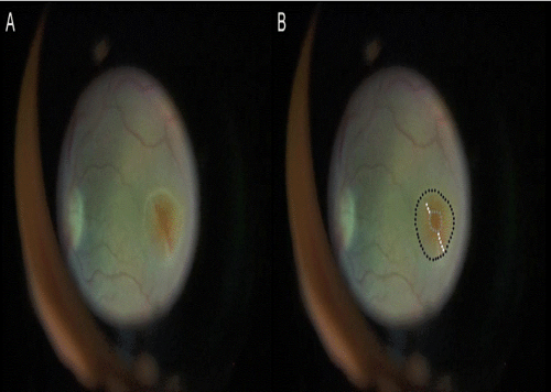
Figure 2:
Macular hole torn inferiorly and superiorly (A). Changes outlined (B); black lines showing localised retinal detachment, white lines showing tears, grey line showing original extent of macular hole.