 |
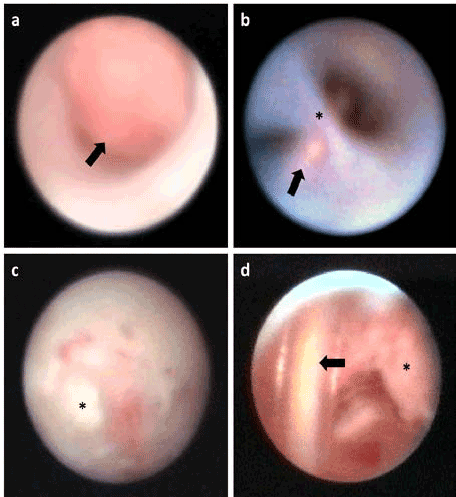
| Figure 2: Endoscopic findings. a) Granulomatous tissue (arrow) of the inferior canaliculus after lacrimal surgery because of trauma, b) View of entrance of the saccus; lacrimal fistula localized by dacryoendoscopy and Bowman probe (arrow) and presaccal stenosis (star), c) Chronic dacryocystitis with fibrinous plaques (star) due to postsaccal re-stenosis after probing and syringing, d) Intrasaccal silicone tube (arrow) causing chronic dacryocystitis with granulomatous tissue (star). |