 |
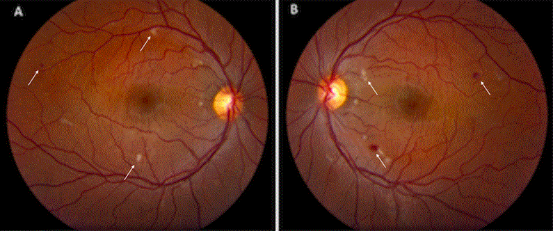
| Figure 1: Fundus photography at time of presentation. A: Fundus photograph of the right eye at presentation depicting cotton wool spots and intraretinal hemorrhages localized to the posterior pole. B: Fundus photograph of the left eye at presentation depicting a similar presentation.c |