 |
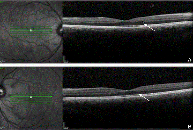
| Figure 2: Optical coherence tomography at time of presentation. A: Spectral-domain optical coherence tomography scan of the right eye at presentation demonstrating gross irregularity of the inner and outer retina with partial loss of the outer plexiform layer. B: Spectral-domain optical coherence tomography scan of the left eye at presentation demonstrating an analogous appearance. |