|
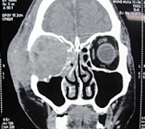
| Figure 3b: Coronal view showing multiple enhancing lesions in superficial lobe of right parotid gland with area of central necrosis& extension of mass lesion into right frontal sinus, ethmoidal air cells with erosion of lamina papyracea and floor of obit. |