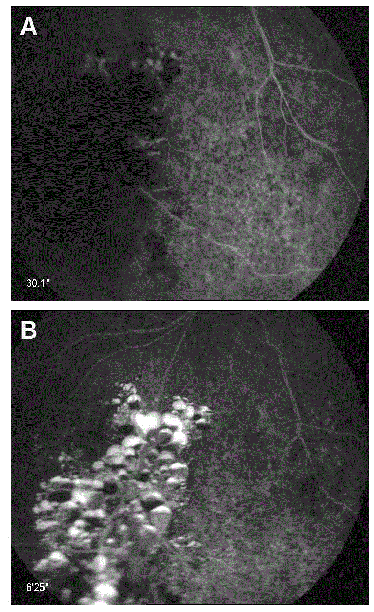
Figure 2:
Fluorescein angiography of a case of retinal cavernous hemangioma. Note hypofluorescence within the tumor in the early phase (A) and hyperfluorescence and incomplete perfusion of the lesion in the late phase (B).