 |
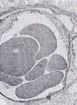
| Figure 4: Microphotography obtained with transmission electron microscopy of a little neo-formed micro-artery within the rabbit cornea after 3 weeks from the alkali-burn. The vessel (diameter<25 µm) contains in the lumen many red cells. On the right side two pericytes can be observed. Magnification=9800X. |