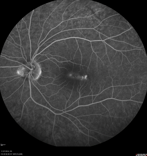
Figure 1c:
Case 1: Fluorescence-angiography-punctate outer retinal toxoplasmosis.