|
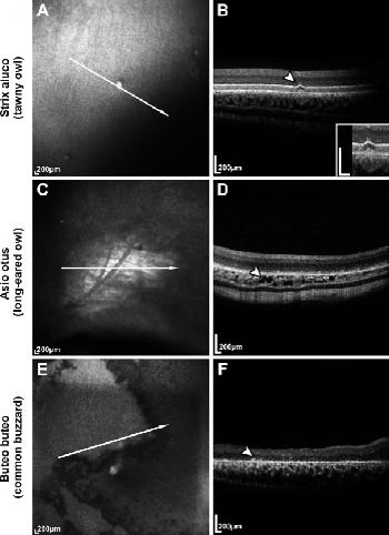
| Figure 3: Images of infrared funduscopy (left column: A, C, E) and of 2D OCT scans (right column: B, D, F) show various morphological and pathological alterations in retinal and subretinal regions in eyes from three different bird species. A and B Bright spots in the fundus image of a tawny owl (Strix aluco) appear as sub-retinal drusenoid changes (arrowhead in B) in 2D OCT image (magnified in the inset). C and D Infrared fundus image and OCT of a long-eared owl (Asio otus) revealed pigmentation abnormalities (visibility of choroidal blood vessels). The arrowhead in D indicates the corresponding area in the 2D OCT image. Related to the depigmented fundus area (C), one might assume washed-out and less distinguishable photoreceptors cell layers and this therefore might represent some evidence for retinal degeneration. E and F The infrared fundus image of a common buzzard (Buteo buteo) shows an extensive pathological altered fundus. 2D OCT reveals distinct atrophic neurodegeneration of the retina and loss and thinning of the retinal layers in the altered fundus areas (arrow head). White arrows in A, C, D indicate the orientation of the corresponding OCT-scan in the right column. |