 |
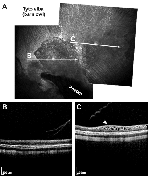
| Figure 4: Arranged image of two adjacent infrared fundus pictures (A) of the right eye of a barn owl (Tyto alba) and the corresponding 2D OCT images (B and C). The white arrows indicate the orientation of the OCT scans. The infrared funduscopy reveals an extensive central area of considerably altered fundus pigmentation near the pecten (white arrow B in A) and a less conspicuous area of changed pigmentation beside it (white arrow C in A). Noticeable, the OCT scans indicate no such obvious pathological changes in the severe altered pigmentation area (B), whereas the less conspicuous area shows clearly disorganization with oedemantous gaps which can be characterized as intra-retinal degenerations (arrowhead in C). |