|
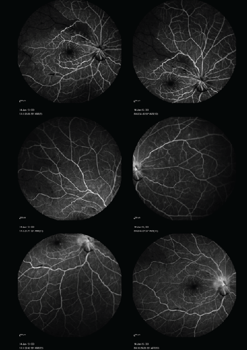
| Figure 2: Right eye. Fluorescein angiography on the day of presentation showing diffuse scattered retinal hypofluorescence due to hemorrhages with tortuous dilated retinal veins in all four quadrants, late disc and mild macular leakage, and no significant non-perfusion area, except non-perfusion along superotemporal arcade over superior macula with delayed branch retinal artery perfusion. |