|
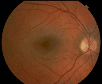
| Figure 9: Fundus photo of the right eye at three months. Fundus photo of the right eye three months after presentation demonstrated residual nasal optic disc edema and minimal hard exudates in a radial pattern around the fovea with an irregular foveal light reflex. |