 |
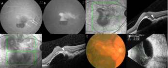
| Figure 2: Ophthalmic examination in case 2. A: Early-phase fluorescein angiography shows a hyperfluorescent area on the macula with blocked fluorescence and multiple brighter spots (arrows). B: Late-phase fluorescein angiography. C: Optical coherence tomography shows pigmented epithelium detachment with hyper-reflectivity localized underneath the dome, raising suspicion for a polypoidal choroidal vasculopathy-associated polyp at the site corresponding to the early brighter spot on fluorescein angiography (arrow head). D: Optical coherence tomography after three monthly injections of ranibizumab with a suspicious polypoidal choroidal vasculopathy-associated polyp (arrow head). E. Fundus photography one month after the first aflibercept injection. F. B-scan ultrasonography six weeks after the first aflibercept injection. |