 |
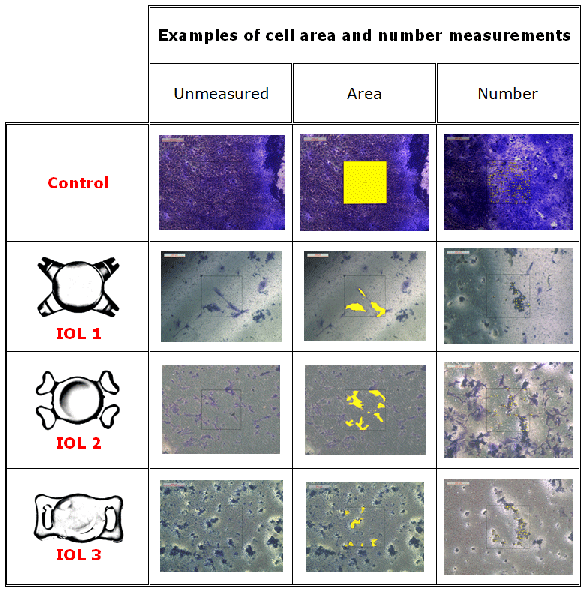
| Figure 4: Examples of measured PET membranes are shown. The area as well as the number of cells was calculated using Leica’s digital microscope software. Every cell covered area and each single cell under the IOL was marked and the total area or total number of cells was calculated by the software. |