 |
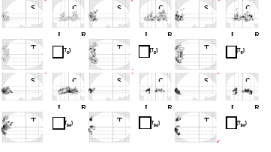
| Figure 3: shows fMRI activation (sagital, coronal and transversal maximum intensity projection) in group of healthy volunteers (upper row) and patient with binasal hemianopia (bottom row) on a), d) bilateral stimulation, b), e) stimulation of the right eye and c), f) stimulation of the left eye. |