|
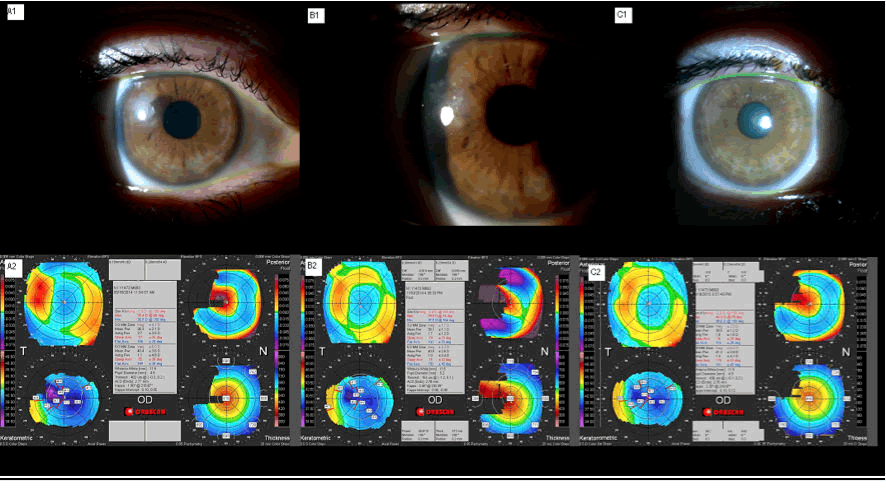
| Figure 4: Clinical and topographic evolution of a case. A1 illustrates grade III epithelial ingrowth; A2: Corneal topography shows 3.5D astigmatism and a flattened profile corresponding to the ingrowth area B1 illustrates the post YAG laser clinical aspect and 2.4D corneal astigmatism, B2. C1 portrays a corneal translucent residual leukoma, one year after YAG laser. C2 shows 2.0D corneal astigmatism and a more regular anterior surface, after one year; mean Km from 38.5 to 39D. |