 |
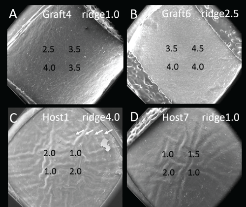
| Figure 6: SEM images at 23X magnification in interface after simulated surgery. In each image, the ridge score is indicated at the upper zone as a white colored value, and the roughness scores are indicated at the 4 quadrants as 4 black colored values. A: A microkeratome-cut anterior lamellar graft with the least ridge (ridge score 1.0). B: A microkeratome-cut anterior lamellar graft with ridge score 2.5. C: The worst ridge score 4.0 with many concentric ridges (arrows) in Host 1. D: A host bed surface with the least ridge (ridge score 1.0) in Host 7. |